


สล็อตเว็บตรง ไม่ผ่านเอเย่นต์ ไม่มีขั้นต่ำ เว็บสล็อต แตกง่าย แตกหนัก อันดับ 1
สล็อตเว็บตรง อันดับต้น ๆ ของเอเชีย สล็อตเว็บตรง ไม่ผ่านเอเย่นต์ ไม่มีขั้นต่ำ เว็บสล็อตสัญชาติไทยมาตรฐานระดับสากล เปิดให้บริการเกมออนไลน์จากหลายค่ายดัง รวมทุกเกมทำเงินไว้ใน เว็บสล็อต ของเราเรียบร้อยแล้ว คุณจะได้พบกับเกม สล็อตแตกง่าย มากกว่า 500 รายการ มาพร้อมระบบภายในเว็บไซต์ที่พัฒนามาเป็นอย่างดี หากคุณกำลังมองหาเกม สล็อตแตกหนัก ไม่ต้องลงทุนเยอะก็รวยได้ เลือกเดิมพันกับเว็บ สล็อตอันดับ 1 ในไทย รับรองว่าเส้นทางการเป็นเศรษฐีของคุณอยู่ใกล้แค่เอื้อม

สล็อตเว็บตรง ไม่ผ่านเอเย่นต์ ไม่มีขั้นต่ำ ระบบออโต้ ฝาก-ถอนไวภายใน 3 วินาที
สล็อตเว็บตรง เปิดให้เดิมพันทุกเกมได้แบบไม่มีขั้นต่ำ สล็อตเว็บตรง ไม่ผ่านเอเย่นต์ พัฒนาระบบฝากถอนใหม่ทั้งหมด ติดตั้งระบบออโต้เพื่อให้สมาชิกทำธุรกรรมทางการเงินได้ง่ายขึ้น เว็บสล็อตไม่มีขั้นต่ำ เลือกที่จะมอบความสะดวกสบายให้กับผู้เล่นเป็นอันดับแรก เพราะเรารู้ดีว่าความสนุกและเงินรางวัล เป็นเรื่องที่รอไม่ได้ เราจึงพัฒนาระบบ สล็อตฝากถอน ไวภายใน 3 วินาที ทำรายการได้เองผ่านระบบ สล็อตออโต้ ผู้เล่นไม่จำเป็นต้องแจ้งแอดมินทำรายการทางการเงินให้อีกต่อไป เพราะทุกขั้นตอนคุณสามารถทำเองได้ ไม่ยุ่งยาก
สล็อตเว็บตรง แตกง่าย แตกหนัก เว็บลิขสิทธิ์แท้ มาแรง อันดับ 1 ทำเงินได้จริง
อย่างที่บอกว่าเราคือ สล็อตเว็บตรง แตกง่าย ชั้นนำของประเทศไทย จึงทำให้มียอดผู้เข้าใช้งานเป็นจำนวนมากทุกวัน เว็บเดียวที่รวมเกม สล็อตแตกง่าย ไว้เยอะที่สุด ทุกครั้งที่ค่ายเกมเปิดตัวเกม สล็อตแตกหนัก ใหม่ล่าสุด เว็บเราจะนำเข้าเกม สล็อตลิขสิทธิ์แท้ ไว้ให้สมาชิกเดิมพันก่อนใครทันที เรียกได้ว่าเป็นเว็บด สล็อตอันดับ 1 ที่ทันสมัย และก้าวทันโลกมากที่สุด เพราะเราไม่ได้นำเข้าเฉพาะเกมใหม่เพียงอย่างเดียว แต่เรายังมีเกมสล็อตทั้งในและนอกกระแส ไว้บริการสมาชิกอีกด้วย ทุกเกม สล็อตได้เงินจริง สมัครสมาชิกกับ เว็บแท้ ครั้งเดียว ใช้ยูสเซอร์ได้ตลอดชีวิต เล่นได้ทุกเกม ไม่ต้องสมัครหลายครั้งให้ยุ่งยาก

สมัคร เว็บสล็อต เว็บตรง ค่ายใหญ่ รับโปรโมชั่นพิเศษมากมาย
สมัครสมาชิกกับ สล็อตเว็บตรง ปุ๊บรับโบนัสพิเศษได้ปั๊บ ไม่ต้องรอลุ้นนาน สมัครสล็อต เว็บตรงอันดับ 1 ลุ้นรับรางวัลพิเศษจากกิจกรรมดี ๆ ได้มากมาย เว็บสล็อต เว็บตรง มาพร้อมข้อเสนอที่น่าสนใจอีกเพียบ เราอัปเดตโปรโมชั่นใหม่สำหรับสมาชิกทุกเดือน คิดค้นโปรโมชั่นเพื่อสมาชิกของ สล็อตค่ายใหญ่ ใหม่ที่สุด โดยเฉพาะ เรามาดูกันว่าเริ่มต้นเดิมพันกับเว็บไซต์ของเรา ผู้เล่นจะได้รับ โปรโมชั่นสล็อต แบบไหนบ้าง ?
• ฝาก 10 รับ 100
สล็อตเว็บตรง แจกโปรโมชั่น ฝาก 10 รับ 100 เอาใจผู้เล่นทุนน้อย และสมาชิกใหม่โดยเฉพาะ ใครจะไปคิดว่าการฝากเงินเข้าระบบแค่หลักสิบ สามารถถอนเงินหลักร้อย นำมาใช้ได้จริง ถ้าคุณเลือกลงทุนกับเว็บไซต์ของเรา คุณสามารถรับข้อเสนอดี ๆ แบบนี้ได้จริง เราแจกให้ฟรีทุกยูสเซอร์ กดรับโปรได้ตลอดทั้งวัน ไม่ต้องทำกิจกรรมให้ยุ่งยาก ถ้าคุณรู้ตัวว่าเป็นนักปั่นทุนน้อย เลือกรับโปรโมชั่นนี้ รับรองว่าไม่ผิดหวัง
• เครดิตฟรี 100
โปรโมชั่นเครดิตฟรี ถือว่าเป็นโปรโมชั่นสุดฮอตที่นักลงทุนให้ความสนใจมากที่สุด เนื่องจากโปรโมชั่น สล็อตเครดิตฟรี สามารถใช้เป็นทุนในการเดิมพันได้จริง ผู้เล่นสามารถกดรับโปรนี้ไปต่อยอดเกมสล็อตได้ทุกค่าย เว็บเราไม่กำหนดเงื่อนไขในการใช้โปรโมชั่น เครดิตฟรี 100 ที่ยุ่งยาก เพียงแค่คุณสมัครสมาชิกกับเว็บเราให้เรียบร้อย หลังจากนั้นก็สามารถล็อกอินเข้าสู่ระบบ เพื่อเริ่มต้นความสนุกได้เลย
• ฝากถอนไม่มีขั้นต่ำ
จะดีแค่ไหน ? ถ้าคุณสามารถเล่นเกมสล็อตได้อย่างอิสระ โดยที่ไม่ต้องกังวลเรื่องเงินทุน เข้าใจถึงปัญหาของผู้เล่นทุนน้อยเป็นอย่างดี เราจึงมอบโปรโมชั่น ฝากถอนไม่มีขั้นต่ำ ให้กับสมาชิกทุกคน โดยเฉพาะผู้เล่นทุนน้อย ที่มีเงินทุนจำกัด คุณสามารถใช้โปรโมชั่น สล็อตฝากถอนไม่มีขั้นต่ำ เพื่อสร้างประสบการณ์ใหม่ ๆ ให้กับตัวเองได้ เรารับรองว่าการเล่นสล็อตของคุณจะสนุกมากขึ้นเป็นสองเท่า!

แนะนำเกม สล็อตเว็บตรง pg โบนัสแตกง่าย เล่นได้จ่ายจริงทุกยอด
คุณกำลังตามหาเกม สล็อตเว็บตรง มาแรง ทำเงินได้จริงอยู่ใช่หรือไม่ ? ถ้าใช่ วันนี้เรามี เกมสล็อต แตกง่าย จากหลากหลายค่ายดังมานำเสนอ เพื่อเป็นแนวทางสำหรับสมาชิกใหม่ ที่ยังไม่รู้จักเกม สล็อตแตกหนัก มากนัก อย่างที่รู้กันดีว่าเกมสล็อตเว็บเรามีให้เลือกเยอะ นำเข้าเกมใหม่ก่อนใครอยู่เสมอ และทุกเกมล้วนมีคุณภาพ โบนัสสล็อต เยอะ แจ็คพอตแตกง่ายทุกเกม ลงทุนแบบไม่มีขั้นต่ำได้ แต่ทำกำไรได้สุดปัง เรามาดูกันว่าเกม สล็อตจ่ายจริง จากเว็บตรงไม่ผ่านเอเย่นต์ มีเกมอะไรบ้าง ?
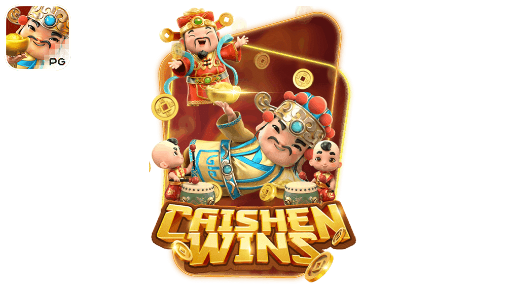
1. เกมสล็อต Caishen Wins สล็อตสายมู
Caishen Wins เกมสล็อตใหม่ล่าสุด ที่เตรียมเงินรางวัลไว้แจกจ่ายสำหรับผู้เล่นมหาศาล เกมนี้เหมาะสำหรับผู้เล่นสายมู เนื่องจากธีมเกมเกี่ยวกับอาแปะ ที่ตามตำนานกล่าวว่าเป็นผู้ให้โชค ประทานพร ให้กับผู้ที่พบเห็น เกมสล็อตแตกง่ายจากค่าย PG slot ที่มาในรูปแบบเกม สล็อตแมชชีน 6 วงล้อ 5แถว ค่า RTP ของเกมอยู่ที่ 96.92% คุณพร้อมรับพรจากอาแปะผู้ศักดิ์สิทธิ์แล้วหรือยัง ? ถ้าพร้อมแล้วตามเรามาได้เลย

2. เกมส์สล็อต Secrets of Cleopatra สล็อตคลีโอพัตรา
เริ่มต้นการเดินทางของคุณไปกับเกมสล็อต Secrets of Cleopatra สาวงามที่เต็มไปด้วยความลับ ภายในเกมคุณจะได้พบกับการตามล่าหาสมบัติ ที่เต็มไปด้วยภัยอันตราย ความงามของคลีโอพัตราส่งผลให้ชายหลาย ๆ คนตกหลุมรัก และกลายเป็นสงครามในที่สุด เกมสุดฮิตจากค่าย พีจีสล็อต เว็บตรง มาในรูปแบบเกม สล็อตออนไลน์ 3 วงล้อ 4 แถว และมีค่า RTP ที่สูงถึง 96.74% นอกจากนี้ยังมีฟีเจอร์พิเศษเป็นตัวช่วยอีกเพียบ

3. เกมสล็อต Treasures of Aztec สล็อตสาวถ้ำ
มาต่อกันที่เกมสล็อตในตำนานอย่าง Treasures of Aztec หรือเกม สล็อตสาวถ้ำ ที่เซียนหลาย ๆ คนยกให้เป็นเกมทำเงินอันดับ 1 ใน สล็อตเว็บตรง ธีมเกมบอกเล่าเรื่องราวเกี่ยวกับประเทศแม็กซิโก และชาวมายัน ที่เป็นต้นกำเนิดของประติมากรรมลึกลับมากมาย มาในรูปแบบเกมสล็อตออนไลน์ 6 วงล้อ 5 แถว รับฟรีสปินได้สูงสุดถึง 10 ครั้ง ฟีเจอร์พิเศษในเกมมีให้เลือกรับอีกเพียบ

4. เกมส์สล็อต Lucky Neko สล็อตแมวเนโกะ
Lucky Neko เกมสล็อตสุดน่ารักจากทางค่ายพีจี รูปแบบเกมมีเอกลักษณ์ รางวัลโบนัสแจ็คพอต มีให้ลุ้นเยอะ ถ้าคุณเป็นแฟนพันธุ์แท้เกม สล็อต PG เว็บตรง แตกหนัก ต้องห้ามพลาดเกม สล็อตแมวเนโกะ เพราะเกมนี้มาพร้อมกับความโชคดีและเงินรางวัลมหาศาล เกมสล็อต 6 วงล้อ 5 แถว ให้อัตราการจ่ายสูงสุดถึง 20,000 เท่าของเงินเดิมพัน รองรับการใช้งานได้ทั้งระบบ IOS และ Android

5. เกมสล็อต Ways Of The Qilin สล็อตกิเลน
Ways Of The Qilin เกมสล็อตที่เนื้อเรื่องเกี่ยวกับสัตว์ในตำนานอย่าง “กิเลน” เนื้อเรื่องในเกมน่าสนใจ ภาพและเสียงถูกสร้างขึ้นอย่างประณีต และพิถีพิถันอย่างที่สุด มาในรูปแบบเกมสล็อตออนไลน์ 6 วงล้อ 6 แถว ให้อัตราการจ่ายสูงสุดถึง 80,000 เท่าของเงินเดิมพัน ค่าย PG เว็บตรง นำเสนอเรื่องราวของสัตว์มงคลอย่างกิเลนได้อย่างสมบูรณ์แบบที่สุด เรียกได้ว่าเป็นอีกหนึ่งเกมสล็อตออนไลน์ภาพสวย ที่คุณไม่ควรพลาด
ข้อดีของ สล็อตเว็บตรง ดีกว่าเว็บอื่นๆอย่างไร
นำเสนอเกมน่าเล่นและการบริการภายในเว็บไซต์บางส่วนไปบ้างแล้ว ต่อไปเรามาดูหัวข้อเกี่ยวกับข้อดีของ สล็อตเว็บตรง กันบ้างดีกว่า เชื่อว่าผู้เล่นหลาย ๆ คน ต่างก็สงสัยว่าเว็บไซต์ของเราดีกว่าเว็บอื่นยังไง ? ทำไมเราถึงถูกยกให้เป็นเว็บสล็อตอันดับ 1 ในไทย ที่มาตรฐานเทียบเท่าเว็บสล็อตต่างประเทศ วันนี้เราได้ทำการรวบรวมข้อดี ๆ จากผู้ใช้งานจริงมานำเสนอให้กับผู้เล่น เพื่อให้คุณตัดสินใจได้ง่ายขึ้น ว่าควรสมัครสมาชิกกับเว็บไซต์ของเราดีไหม ซึ่งข้อดีหลัก ๆ ของทางเว็บไซต์มีดังนี้
• มีโอกาสในการชนะที่เยอะกว่า
เกมสล็อตเว็บตรงเพิ่มอัตราการแตกของรางวัลแจ็คพอตให้มากขึ้น นั่นหมายความว่าคุณสามารถลุ้นรับรางวัลแจ็คพอตได้มากขึ้น ถ้าเลือกเดิมพันกับเว็บเรา เดิมทีเรานำเข้าเกมสล็อตแตกง่ายจากทุกค่ายชั้นนำอยู่แล้ว ยิ่งเราพัฒนาระบบให้มากขึ้น โอกาสในการลุ้นรางวัลแจ็คพอตก็ยิ่งมากขึ้นตามไปด้วย คุณสามารถสร้างตัวจากเกมสล็อตเว็บเราได้จริง เพียงแค่คุณต้องฉลาดเลือกเกมที่เหมาะกับตัวเอง และลงทุนอย่างมีสติ
• เป็นเว็บที่ใช้งานง่ายและสะดวก
ถ้าคุณเป็นผู้เล่นสายเทคโนโลยี ชอบความทันสมัย เลือกเดิมพันกับเว็บเรา รับรองว่าตอบโจทย์ เพราะเว็บเราใช้งานง่าย ล็อกอินเข้าสู่ระบบได้อย่างสะดวกสบายผ่านมือถือทุกรุ่น เท่านั้นยังไม่พอ เรายังติดตั้งระบบอัตโนมัติไว้อำนวยความสะดวกให้กับผู้เล่นอีกด้วย เรียกได้ว่าเป็นเว็บไซต์ที่ครบวงจรในทุก ๆ ด้าน พร้อมพัฒนาระบบอย่างต่อเนื่อง อัปเดตทางเข้าใช้งานให้ทันสมัยอยู่เสมอ เพื่อให้ตรงใจสมาชิกมากที่สุด
• มีการรักษาความปลอดภัยสูง
ถ้าไม่อยากโดนโกงให้เลือกลงทุนกับสล็อตเว็บตรง เพราะความปลอดภัยค่อนข้างแน่นหนา รักษาความเป็นส่วนตัวให้กับสมาชิกได้เป็นอย่างดี เดิมพันกับเว็บไซต์ของเราไม่ต้องกลัวว่าข้อมูลส่วนตัวจะรั่วไหล หรือเงินหายจากบัญชี เว็บเราเน้นความปลอดภัยของสมาชิกเป็นหลัก ฝากเงินไว้เท่าไหร่ถอนคืนได้เท่านั้น ไม่มีการหักค่าธรรมเนียม สมัครสมาชิกได้เองผ่านระบบอัตโนมัติ ไม่จำเป็นต้องแจ้งข้อมูลส่วนตัวกับบุคคลที่สาม
• มีโปรโมชั่นแจกเยอะ
ถ้าคุณเป็นผู้เล่นสายล่าโปร ชอบความคุ้มค่า เลือกลงทุนกับเว็บเรา คุณจะได้ทุกสิ่งที่คุณต้องการ สล็อตเว็บตรงดีลโปรเด็ดโดนใจตลอดทั้งเดือน กดรับโปรง่าย ไม่ต้องฝาก ไม่ต้องแชร์ ให้ยุ่งยาก เพียงแค่คุณสมัครสมาชิกกับเรา และยืนยันตัวตนให้เรียบร้อย เพียงเท่านี้ก็สามารถเลือกรับโปรโมชั่นที่สนใจ นำไปต่อยอดเกมสล็อตได้ตามต้องการ ยูสเดียวรับได้หลายโปรโมชั่น เราแจกโปรดีทุกวัน ไม่เว้นวันหยุดราชการ
• การบริการแบบมืออาชีพ
สล็อตเว็บตรงไม่ได้พัฒนาแค่ระบบภายในเพียงอย่างเดียว แต่เรายังพัฒนาบุคลากรของเราอีกด้วย หลังจากสมัครสมาชิกกับเว็บเราเรียบร้อยแล้ว คุณจะได้รับการบริการระดับวีไอพีตลอดการใช้งาน เรามีทีมงานที่มากด้วยประสบการณ์คอยให้คำปรึกษาในทุก ๆ ด้าน ทีมงานของเราได้รับการอบรมมาเป็นอย่างดี แก้ไขปัญหาให้กับผู้เล่นอย่างตรงจุด ดูแลลูกค้าด้วยความเต็มใจ ยิ้มแย้มแจ่มใส ไม่เหวี่ยง ไม่วีน
• ตัวเกมมีประสิทธิภาพสูง ภาพสวย สมจริง
ถ้าคุณเคยเดิมพันเกมสล็อตเว็บอื่น แล้วไม่ประทับใจคุณภาพของตัวเกม เล่นไปเกมกระตุกไป ทำให้การเดิมพันไม่ราบรื่นเท่าที่ควร เปลี่ยนมาเดิมพันกับเว็บเรา คุณจะได้พบกับเกมสล็อตภาพสวยสมจริงทุกอณู เอฟเฟกเน้นความอลังการ เสียงประกอบในเกมช่วยเพิ่มอรรถรสระหว่างเดิมพันให้กับผู้เล่นได้เป็นอย่างดี เป็นการสร้างรายได้ที่มาพร้อมความสนุก ไม่น่าเบื่อ ไม่จำเจ มีให้เลือกเดิมพันทั้งเกมเก่าและเกมใหม่

สล็อตแตกหนัก แตกง่าย เว็บตรง เล่นได้ทุกที่ทุกเวลา ตลอด 24 ชม.
สล็อตเว็บตรง เปิดให้บริการทุกวัน ไม่มีวันหยุด เบื่อไหมเวลาหยุดยาวแล้วไม่รู้จะทำอะไรดี จะไปเที่ยวก็กลัวเปลืองเงิน แดดร้อนบ้าง ฝนตกบ้าง อากาศไม่เป็นใจ ก็ไม่พ้นนอนเหงาอยู่บ้าน สำหรับใครที่วันหยุดไม่รู้จะทำอะไร ไม่รู้จะไปไหนดี เลือกเล่นเกม สล็อตแตกหนัก อยู่ที่บ้านรับรองว่าสนุกไม่แพ้กิจกรรมอื่น เกม สล็อตแตกง่าย ทำเงินได้จริง ดีกว่าใช้เวลาไปกับเรื่องไร้สาระไปวัน ๆ เลือกหารายได้เสริมกับเกมสล็อตจาก เว็บตรง ไม่ผ่านเอเย่นต์ ได้ทั้งความสนุก ได้ทั้งเงินรางวัล เป็นการตอบแทน

เว็บสล็อต แตกง่าย เว็บแท้จากต่างประเทศ มาตรฐานระดับสากล
ถ้าใครเคยใช้บริการเว็บสล็อตต่างประเทศ จะรู้ดีว่ามาตรฐานของเขาน่าประทับใจมากแค่ไหน ทุกขั้นตอนเต็มไปด้วยความรวดเร็ว ไร้ซึ่งข้อผิดพลาดใด ๆ วันนี้คุณสามารถใช้งานกับ สล็อตเว็บตรง มาตรฐานเดียวกันกับเว็บ สล็อตต่างประเทศ ได้แล้ว เรารับรองความประทับใจตั้งแต่ใช้งานครั้งแรก คุณจะได้รับการบริการระดับวีไอพีตลอดการใช้งาน เว็บสล็อต แตกง่าย ไม่มีเด้ง ไม่มีค้าง เดิมพันได้อย่างลื่นไหลทุกเกม ถ้าคุณไม่อยากเสี่ยงเจอเว็บเอเย่นต์ที่ไม่ได้คุณภาพ ให้เว็บเราเป็นคำตอบสุดท้าย รับรองว่าไม่ผิดหวัง
สรุป สล็อตเว็บตรง
การเลือกเดิมพันกับ สล็อตเว็บตรง มาตรฐานสากล เป็นสิ่งที่การันตีรายได้ให้กับผู้เล่นได้มากที่สุด ขั้นตอนการสร้างรายได้จากเกม เว็บตรงสล็อต ไม่อยากเลย เพราะเราคัดเฉพาะเกมสล็อตแตกง่าย ไว้บริการลูกค้าอยู่แล้ว สิ่งที่ สล็อตเว็บตรง ไม่ผ่านเอเย่นต์ ไม่มีขั้นต่ำ เพิ่มเติมให้กับสมาชิกคือ ความใส่ใจ เงินรางวัล โปรโมชั่น และโบนัสพิเศษต่าง ๆ ซึ่งเรารับรองว่าทุกคนจะได้รับสิ่งนี้อย่างเท่าเทียม ไม่ว่าคุณจะลงทุนกับ เว็บตรงสล็อต ไม่ผ่านเอเย่นต์เป็นเงินจำนวนเท่าไหร่ เราก็พร้อมดูแลคุณอย่างเต็มที่ เพราะเป้าหมายของเราคือ การทำความฝันของสมาชิกให้เป็นจริง สร้างรายได้ที่มั่นคงให้กับสมาชิกทุกคน เพื่อเป็นการตอบแทนลูกค้า
FAQ : คำถามที่พบบ่อย
Q: สล็อตเว็บตรง คืออะไร
A: สล็อตเว็บตรงคือการผจญภัยในโลกของเกมสล็อตที่ผู้เล่นสามารถสนุกไปกับมันได้โดยตรงผ่านทางเว็บไซต์ของผู้ให้บริการ ทำให้คุณสามารถสนุกกับเกมอย่างไม่ยากลำบากและได้อย่างรวดเร็ว
Q: สล็อตเว็บตรงมีวิธีการทำธุรกรรมการเงินอย่างไร
A: เรามีระบบฝาก-ถอนที่รวดเร็วและสะดวกสบายสำหรับการจัดการเงินของผู้เล่น เพื่อให้ทุกการทำธุรกรรมเป็นไปอย่างราบรื่น
Q: สล็อตเว็บตรง ไม่ผ่านเอเย่นต์ มีโปรโมชั่นและโบนัสใดบ้าง?
A: มีโปรโมชั่นและโบนัสที่หลากหลายที่ทำให้เกมสล็อตเว็บตรงน่าสนใจ เช่น โปรโมชั่นสำหรับผู้สมัครใหม่, โปรโมชั่นการฝากเงิน, และโปรโมชั่นพิเศษอื่น ๆ เพื่อเพิ่มความน่าสนใจในการเล่น
Q: สล็อตเว็บตรง แตกง่าย มีวิธีเพิ่มโอกาสในการทำให้เกมแตกมากขึ้นได้อย่างไร?
A: เกมสล็อตเว็บตรงที่แตกง่ายมักใช้เทคโนโลยี RNG (Random Number Generator) ที่ถูกต้องและเชื่อถือได้ เพื่อให้ผลลัพธ์ของการหมุนสล็อตเป็นสุ่มและไม่สามารถทำนายได้ล่วงหน้า
Q: สล็อตเว็บตรง อันดับ 1 มักมีคุณสมบัติพิเศษอะไรบ้าง?
A: สล็อตเว็บตรง อันดับ 1 มักมีคุณสมบัติพิเศษ เช่น กราฟิกที่สวยงาม, ระบบเสียงที่ทันสมัย, และเกมที่ทันสมัยที่สร้างประสบการณ์เล่นที่น่าตื่นเต้นและน่าสนใจ